Doctors develop new way to use MRI to predict pregnancy complications
University of California - Los Angeles Health News Dec 15, 2017
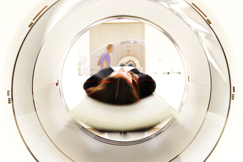
UCLA scientists have developed a new way to use MRI to scan the placenta. The noninvasive approach offers valuable insights into how the mother’s blood enters the placenta and sustains the fetus with oxygen and nutrients during early pregnancy. The technique breaks new ground because most previous studies on this subject occurred in the laboratory after childbirth.
The placenta is a temporary organ that joins a pregnant woman to her baby through the umbilical cord. Few methods exist for safely and accurately studying the placenta in early pregnancy. Ultrasound indirectly measures uterine blood flow, but it is not very effective at predicting pregnancy complications. Another procedure, chorionic villus sampling, can be used to biopsy the placenta, but the test is invasive and can increase the risk of miscarriage and infection.
The researchers used MRI to track water molecules in the arterial blood of 34 women in their second trimester of pregnancy. The new technique allowed the team to measure how much blood entered the placenta and reached the developing baby in the womb, expanding doctors’ ability to accurately determine the health of the placenta.
Using MRI enables doctors to measure blood flow to the placenta and distinguish between normal and abnormal placental function. The information allows them to predict and prevent complications later in pregnancy before symptoms or testing occur. Common complications include premature birth, placental stroke, intrauterine growth restriction and gestational hypertension, and preeclampsia.
The study’s authors are Kyung Sung, assistant professor of radiology; Dr. Carla Janzen, associate professor of obstetrics-gynecology; and Dr. Sherin Devaskar, physician-in-chief at UCLA Mattel Children’s Hospital—all of the David Geffen School of Medicine at UCLA.
The study was published in the Journal of Magnetic Resonance Imaging.
The study was funded by the National Institute of Child Health and Human Development, through its Human Placenta Project.
-
Exclusive Write-ups & Webinars by KOLs
-
 Daily Quiz by specialty
 -
 Paid Market Research Surveys
 -
Case discussions, News & Journals' summaries
